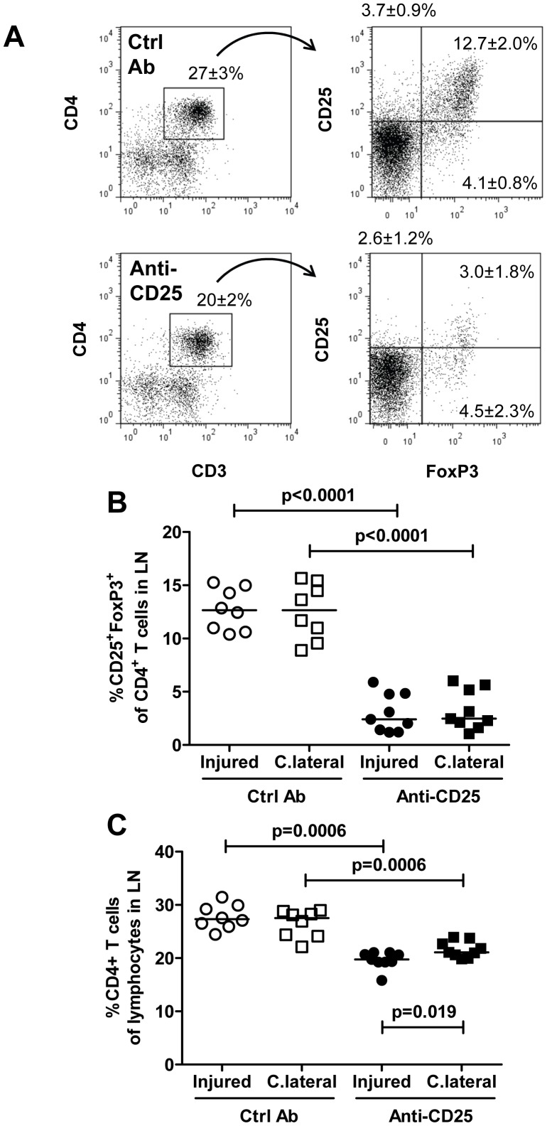
Figure 7. Reduced regulatory T cells in draining lymph nodes after injury of the carotid artery and treatment with anti-CD25.
Cells were isolated from pooled lymph nodes (LN) of injured or sham-operated mice, stained with antibodies against CD3, CD4, FoxP3 and CD25 and analyzed by flow cytometry. A. Representative dot plots. B. FoxP3+ CD25+ cells as a percentage of CD3+CD4+ T cells and C. CD4+ T cells of lymphocytes in lymph nodes draining injured and uninjured contralateral carotid arteries. C.lateral, contralateral; Ctrl Ab, control antibody.

