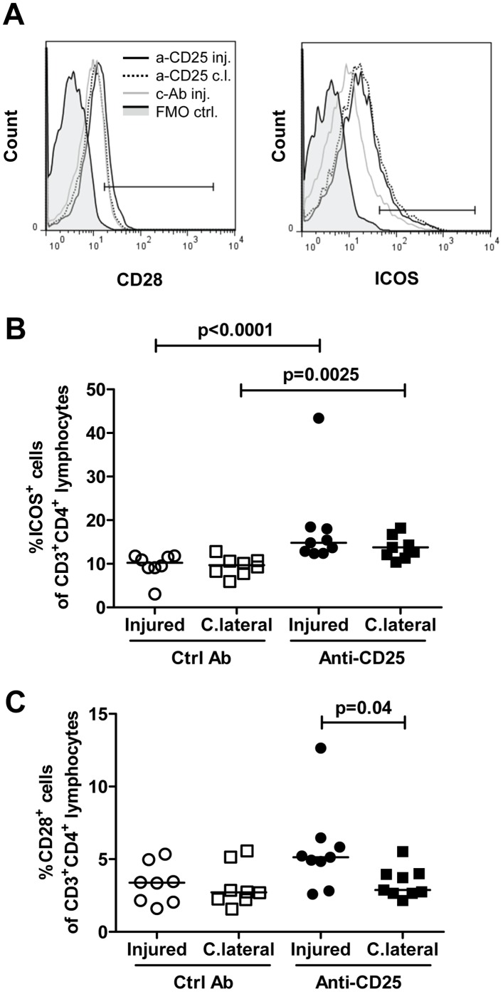
Figure 8. Increased CD28+ and ICOS+ T cells in draining lymph nodes after injury of the carotid artery and blockade with anti-CD25.
Cells were isolated from draining lymph nodes of injured or uninjured contralateral carotid arteries, stained with antibodies against CD3, CD4, CD28 and ICOS and analyzed by flow cytometry. A. Representative histograms. Gate boundaries were set by fluorescence minus one controls (FMO ctrl, solid grey). B. ICOS+ cells as a percentage of CD3+CD4+ T cells. C. CD28+ cells as a percentage of CD3+CD4+ T cells. C.lateral and c.l., contralateral; Ctrl Ab and c-Ab, control antibody; inj, injured.

