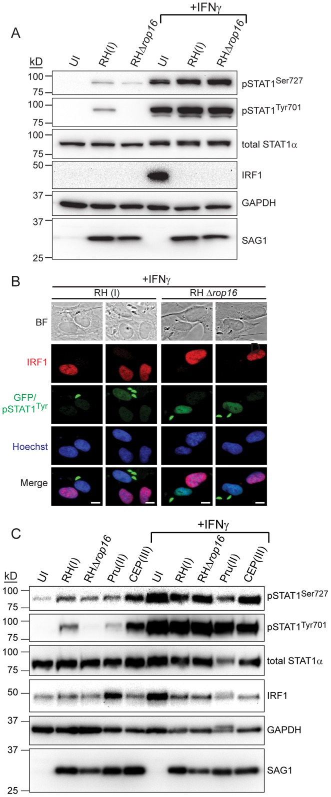
Figure 3. ROP16-activated STAT1 is not transcriptionally active. A.

HFFs were infected with RH(I) or RHΔrop16 parasites at an MOI ∼7, or left uninfected, for four hours. Cells were stimulated, or not, with 100 U/ml human IFNγ for the last hour of infection and cell lysates were collected, run on an SDS-PAGE gel, and Western blotted for phospho-STAT1Ser727, phospho-STAT1Tyr701, total STAT1α, IRF1, GAPDH (host cell loading control) and SAG1 (parasite loading control). This experiment has been performed three times with similar results. B. HFFs were infected with RH(I) or RHΔrop16 parasites for four hours, stimulated with 100 U/ml IFNγ for the last two hours of infection, fixed, and stained with anti-IRF1 (red), anti-phospho-STAT1Tyr (green), and Hoechst dye (nucleus, blue). Parasites also express GFP (green). Scale bar represents 10 µm. This experiment was performed three times with similar results. C. HFFs were infected with RH(I), RHΔrop16, Pru(II), or CEP(III) parasites at an MOI ∼1, or left uninfected, for four hours. Cells were stimulated, or not, with 100 U/ml human IFNγ for the last hour of infection and cell lysates were collected, run on an SDS-PAGE gel, and Western blotted for phospho-STAT1Ser727, phospho-STAT1Tyr701, total STAT1α, IRF1, GAPDH (host cell loading control) and SAG1 (parasite loading control). This experiment has been performed two times with similar results.
