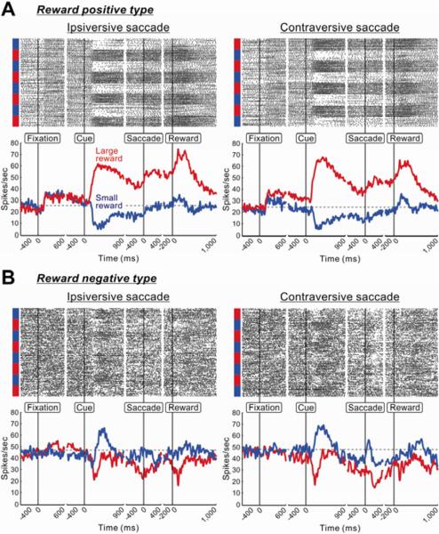
Figure 2.
Two VP neurons showing distinct reward modulations
(A) Spike activity of a neuron showing positive reward modulation. Its activity is shown separately for the ipsiversive (left panel) and contraversive (right panel) saccades in the reward-biased memory-guided saccade task. For each saccade direction, rasters of spikes (top panel) and spike density functions (SDFs, σ = 10 ms; bottom panel) are aligned at the onsets of fixation point, target cue, saccade, and reward delivery. The spike rasters are shown in order of occurrence of trials from top to bottom. Large- and small-reward trials are indicated, respectively, by red and blue bars on the left side of the rasters. The SDFs are shown separately for large-reward trials (red) and small-reward trials (blue); the first trial in each block was excluded. A gray dotted line indicates the baseline firing. (B) Spike activity of a neuron showing negative reward modulation.
Tachibana et al.

