
Figure 3.
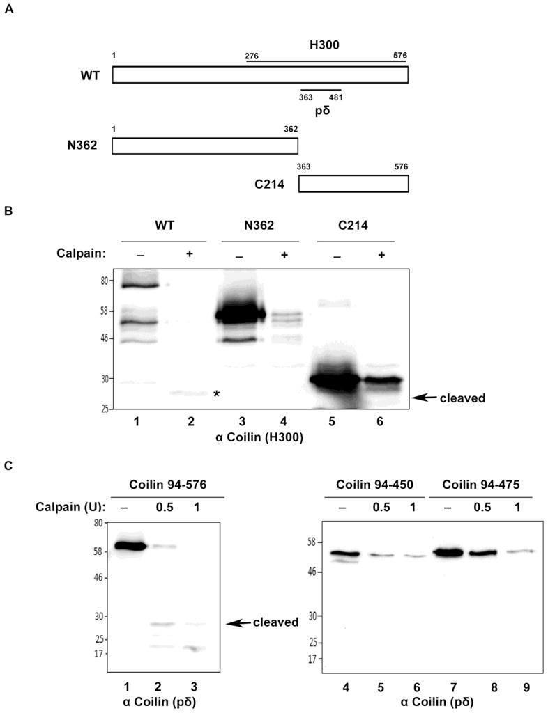
Mapping of coilin digestion by calpain. (A) Schematic representation of full length coilin (WT) and N-terminal (N362) and C-terminal (C214) coilin fragments. The location of the epitopes to which the anti-coilin antibodies H300 and pδ react are shown. (B) Full length coilin, but not N362 or C214, releases cleaved coilin fragment. His-tagged WT, N362 and C214 were incubated with or without calpain in presence of 1mM CaCl2 at 30°C for 15 minutes. The reactions were subjected to SDS-PAGE, Western transfer, and blots probed with anti-coilin H300 antibodies. Asterisk (*) indicates the cleaved coilin fragment. (C) Coilin 94–576 releases the 28 kDa. cleaved coilin fragment. His-tagged coilin constructs containing amino acids 94–576, 94–450 or 94–475 were incubated with 0.5 or 1 U calpain in presence of 1mM CaCl2 at 30°C for 15 min. Reactions were subjected to SDS-PAGE, Western transfer, and blots probed with anti-coilin pδ antibodies.