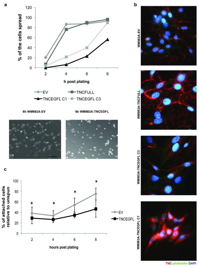
Figure 4. WM983A-TNCEGFL cells present impaired cell spreading and attachment that is TNCEGFL dose dependant.
(A) Cells were imaged at the indicated times after seeding by phase contrast microscopy and scored for the percentage of spread cells (upper panel). Lower panel shows the difference in cell spreading at 6h. Shown are representative of three experiments. Scale bar 100μm. (B) Immunostaining of TNC in two clones of WM983A-TNCEGFL (C1 and C3) and the full length TNC (TNCFULL) construct. Arrowheads point to TNC fibers. (C) WM983A-TNCEGFL detached to a greater extent in inverted centrifugation assays. Shown is an average ± SEM of three experiments, each in triplicate. * p < 0.05.

