Figure 1.
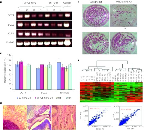
Generation of human induced pluripotent stem (hiPS) cell lines. (a) PCR showing the presence of transgenes (OCT4, SOX2, KLF4, and C-MYC) in hiPS clones generated from human MRC5 and BJ fibroblast cell lines. (b) Alkaline phosphatase staining of hiPS (BJ and MRC5) and wild-type human ES (H1 and H7) colonies. (c) Graphical representation of relative gene expression of pluripotency markers (OCT4, SOX2, and NANOG) from both hiPS (BJ and MRC5) and wild-type human embryonic stem (hES) (H1 nd H7) cell lines. Bars, standard error of n = 3 experiments. (d) Hematoxylin and eosin (HE) staining of teratoma sections obtained from mice 4–6 weeks following subcutaneous injection with 1 × 10⁶ hiPS cells. These sections revealed tissues belonging to the three germ layers (endoderm, mesoderm and ectoderm). Bar = 500 µm. (e) Microarray analysis illustrating closeness between the various wild-type hES and hiPS samples. The correlation coefficient between different wild-type hES cells (H1 and H9) is shown to be 0.9122, which is similar to the correlation coefficient of 0.9164 between hiPS and hES lines.

