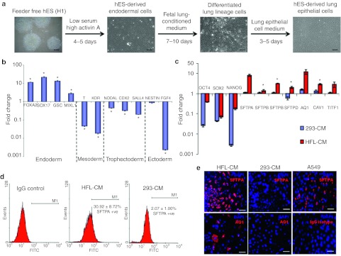
Figure 2.
In vitro differentiation of human embryonic stem (ES) cells toward lung epithelial cells via definitive endoderm. (a) Schematic diagrams summarizing the differentiation protocol adopted in this study. The various media used and representative images of the cellular phenotypes at different stages of differentiation were as shown. Scale = 200 µm. (b) Graphical representation illustrating that human ES cells cultured in low serum, high Activin A medium expressed high levels of endodermal markers after 5 days. Quantitative-PCR on cells after treatment revealed high expressions of endodermal markers such as SOX17, FOXA2, and GSC, but downregulation of other lineage markers. The fold change in gene expression values were normalized to ES cells that were cultured in mouse embryonic fibroblast conditioned medium (MEF-CM). Bars, SD of n = 3 experiments. *P < 0.05 for Kruskal–Wallis one-way analysis of variance compared to control (ES cells cultured in MEF-CM). (c) Quantitative-PCR comparing gene expression of lung specific genes in human embryonic stem (hES)-derived lung epithelial cells (hES-LEC) that were differentiated with either human fetal lung conditioned medium (HFL-CM) or nonspecific medium (293-CM). The fold change in gene expression values were normalized to ES cells that were cultured in MEF-CM. Bars, SD of n = 3 experiments. *P < 0.05 for Kruskal–Wallis one-way analysis of variance compared to control. (d) Flow cytometric analysis of surfactant protein A (SFTPA) positive cells differentiated with either HFL-CM or 293-CM. Human ES cells that have been subjected to the step-wise differentiation process using either HFL-CM or 293-CM was stained with rabbit IgG primary antibody (Isotype control) or rabbit anti-surfactant protein A primary antibody, followed by goat anti-rabbit IgG Alexa 488 (FITC). The percentages of SFTPA-positive cells were as shown. SD of n = 4 experiments. (e) Immunocytochemistry of lung epithelial cells (LECs) differentiated with either HFL-CM or 293-CM. Representative fluorescent images of SFTPA (red) staining of A549 cell line (positive control) and differentiated cells were as shown. Cells differentiated with HFL-CM were also shown to be positive for aquaporin 1 (AQ1). SFTPA (red) was demonstrated to be a cytoplasmic protein with rabbit anti-surfactant protein A primary antibody from Chemicon International, while AQ1 was revealed with rabbit anti-aquaporin 1 (Chemicon International) primary antibody. Cell nuclei (blue) were stained with 4′, 6-diamidino-2-phenylindole (DAPI). Bar = 100 µm.

