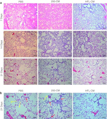
Figure 3.
Time-course study showing lung repair in mice. A total of 27 mice, with 9 mice in each group, were transplanted with phosphate-buffered saline (PBS), 293-CM differentiated cells or human fetal lung conditioned medium (HFL-CM)-differentiated cells, which were derived from human embryonic stem (hES) cells. The recovery process was traced for 21 days at 7 days interval. Lungs from three mice were harvested for analysis at each time point. However, for mice that were transplanted with PBS or with cells that were differentiated with 293-CM, due to availability of surviving mice, only 1 animal was harvested on day 14 and day 21, respectively. (a) Hematoxylin and eosin (HE) staining of lung sections. Lung sections obtained from mice transplanted with PBS, 293-CM differentiated cells and HFL-CM differentiated cells after bleomycin (BLM)-induced lung injury. Note the enlarged air spaces in the lung sections obtained from PBS and 293-CM controls after 21 days. Bar = 200 µm. (b) High magnification of HE staining of lung sections 21 days after cellular transplantation. Enlarged air spaces (yellow arrows) were observed in both PBS control and experimental group transplanted with 293-CM differentiated cells. Thickened alveoli walls were also observed in animal transplanted with 293-CM differentiated cells (red arrows) compared to animals transplanted with HFL-CM differentiated cells. Bar = 400 µm.

