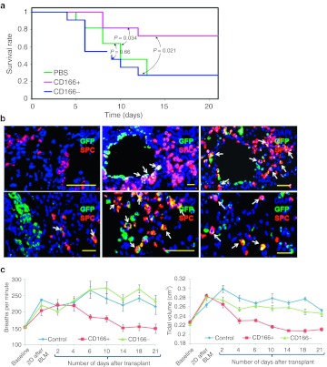
Figure 6.
CD166pos lung epithelial cells (LECs) were responsible for the repair of acute lung injury. (a) Kaplan–Meier curve showing survival rates of mice transplanted with phosphate-buffered saline (PBS), CD166pos or CD166neg LECs derived from human embryonic stem (hES) over a 3 weeks period after bleomycin-induced lung injury. The experiment was performed twice with eight mice in each of the experimental groups. The survival analysis was performed using Kaplan–Meier estimator. The P values between the experimental groups are as shown. (b) Immunocytochemistry of lung section from surviving mice transplanted with CD166pos LECs. Top left panel represents control lung section obtained from surviving mice that were treated with bleomycin, but without cellular transplantation. For mice that were transplanted with CD166pos LECs, the cells were present at the terminal bronchioles and appeared as clusters in the alveolar regions of damaged areas. Some of the green fluorescent protein (GFP)-positive cells were shown to be positive for SPC, a specific marker for type II pneumocyte (white arrow). Clusters of GFPpos CD166pos LECs eventually spread out to form new alveoli. Cell nuclei (blue) were stained with 4, 6-diamindino-2-phenylindole (DAPI). Sections were scanned with a high-resolution MIRAX MIDI system (Carl Zeiss) equipped with both bright field and fluorescence illumination. Images were then analyzed by the software Miraxviewer. Bar = 50 µm. (c) Measurement of lung pulmonary functions was performed by whole body plethysmography of mice transplanted with PBS, CD166pos or CD166neg lung epithelial cells (LECs). The breathing rates and tidal volumes of the three groups of mice were monitored over a 3 weeks period. Error bars represent standard deviation obtained from surviving mice at each time point.

