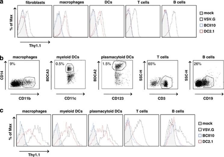
Figure 3.
Selective transduction of human DCs and macrophages by DC2.1 displaying LVs. (a) Human fibroblasts, blood-derived B and T cells, in vitro generated macrophages and bone marrow-derived DCs were mock transduced or transduced with VSV.G pseudotyped, or Nb BCII10 or DC2.1 displaying LVs (multiplicity of infection 10). Flow cytometry was performed 72 h later to evaluate transgene (Thy1.1) expression. The histograms demonstrate Thy1.1 positivity in the evaluated cell types. The tinted, blue, black and red histogram, represent mock transduced cells or cells transduced with Nb BCII10 displaying LVs, VSV.G pseudotyped LVs and Nb DC2.1 displaying LVs, respectively. One representative experiment is shown (n=3). (b, c) Single cell suspensions prepared from human LNs were transduced in vitro with Thy1.1 encoding LVs (multiplicity of infection 10). In order to evaluate Thy1.1 expression in macrophages, myeloid DCs, pDCss, B and T cells, these cells were co-stained with the anti-Thy1.1 antibody and antibodies directed against CD11b CD14, CD11c BDCA-3, CD123 BDCA-2, CD19 and CD3, respectively. The flow cytometry graphs in panel C depict Thy1.1 expression; the corresponding cell populations are shown in the histograms displayed in panel (b). The tinted, blue, black and red histogram represent, mock transduced cells or cells transduced with Nb BCII10 displaying LVs, VSV.G pseudotyped LVs or Nb DC2.1 displaying LVs, respectively. One representative experiment is shown (n=3).

