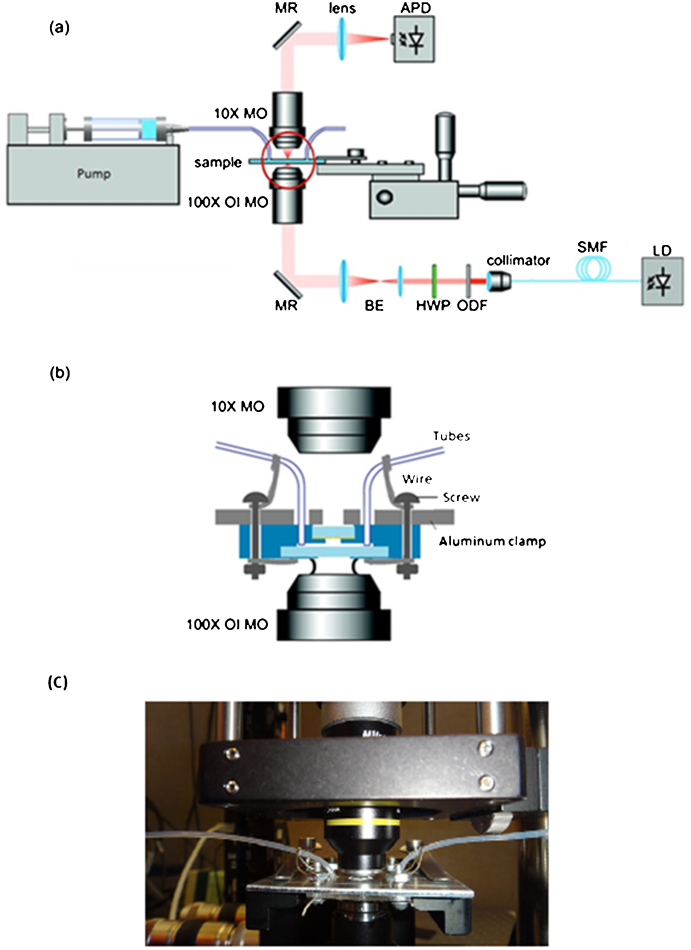
Figure 1.
(a) Schematic drawing of the nanoscale double-hole optical trap. (b) An enlargement of the red circle in part (a), showing details of the composition of the sample in the microfluidic chip, the setup of the oil immersion microscope objective, and the condenser microscope objective. (c) Photograph of the working setup. Abbreviations used: LD = laser diode; SMF = single-mode fiber; ODF = optical density filter; HWP = half-wave plate; BE = beam expander; MR = mirror; MO = microscope objective; OI MO = oil immersion microscope objective; APD = avalanche photodetector.

