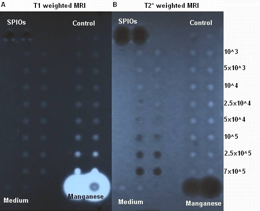
Figure 1.

1T MRI scan of MnCl2and SPIOs labelled cells in a 1% agar phantom. The numbers of labelled cells accounted: 7 × 10⁵, 2.5 × 10⁵, 105, 5 × 10⁴, 2.5 × 10⁴, 104, 5 × 10³, 103. As controls 1 × 10⁵ unlabelled cells, the culture medium, 1 M MnCl2, and Endorem solution were used. A: T1 weighted MRI. SPIOs labelled cells (left two lanes), the controls with un-labelled cells and the culture medium showed comparable signals. 1 M MnCl2 solution showed a strong signal enhancement. MnCl2 labelled cells (right two lanes) were detected to a limit of 105 cells. B: T2* weighted MRI scan. MnCl2 labelled cells (right two lanes), the controls, and the culture medium showed comparable signals. Endorem solution showed a strong signal extinction. SPIOs labelled cells (left two lanes) were detected to a limit of 105cells.
