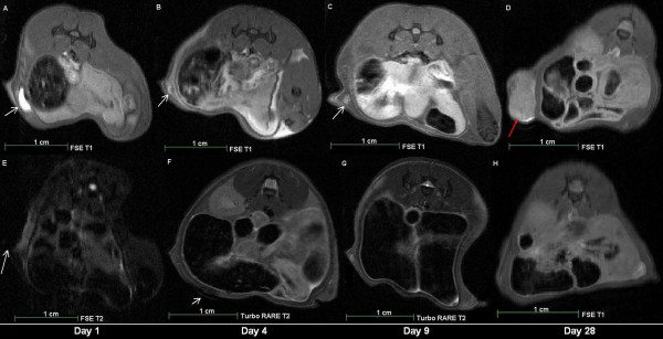
Figure 3.
7T MRI scans of a male NOD-scid mouse after subcutaneous injection of 104SPIOs labelled CT1258 cells. The MRI scans were performed on the same day as the injection (day 1). A: FSE T1 weighted MRI Scan. B: FSE T2 weighted MRI scan. C: FISP T2* weighted MRI scan. Arrows: localisation of the injected SPIOs labelled cells in T1 (A), T2 (B) and T2* (C) weighted MRI. In T2 (B) and T2* (C) weighted MRI the SPIOs labelled cells were detected due to a strong signal extinguishment.

