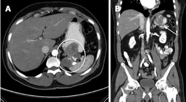
Figure 4.

Locally-advanced gastric gastrointestinal stromal tumors. A: Representative contrast-enhanced computed tomography images show a large, proximal gastric gastrointestinal stromal tumors that invades into the splenic hilum (oval); B: On the coronal images the arrow indicates a heterogeneous mass invading into the spleen with areas of viable tumor and necrotic areas represented by calcifications.
