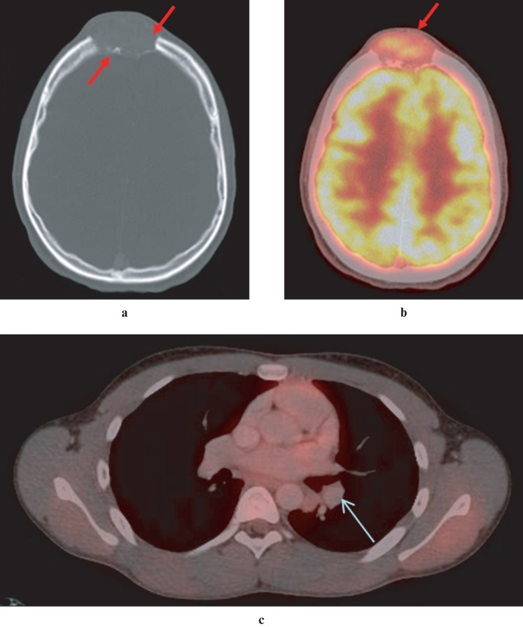
Figure 4.
(a) Axial CT image demonstrates an expansile, osteolytic lesion, eroding the outer and inner tables of the frontal bones in the midline. (b) Axial positron emission tomography (PET)-CT image demonstrates moderate 18-fluorodeoxyglucose (FDG) uptake (maximum standardized uptake value (SUVmax) 6.0) in frontal soft tissue mass, suggestive of malignancy. Intense FDG uptake in the grey matter of the brain is physiological. (c) Axial PET-CT image of the thorax demonstrates moderate FDG uptake (SUVmax 2.0) in a 12 mm × 9 mm nodule in the lower lobe of the left lung, suggestive of pulmonary metastasis

