
Figure 5.
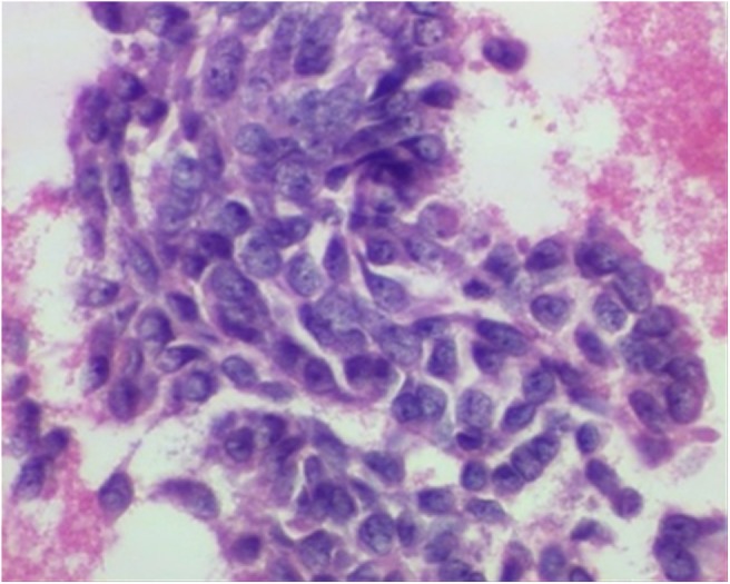
High-power microscopic image of haematoxylin and eosin stain of a specimen from a fine needle aspiration of the skull lesion demonstrates densely packed blue spindle cells consistent with metastatic ameloblastic carcinoma
Official websites use .gov
A
.gov website belongs to an official
government organization in the United States.
Secure .gov websites use HTTPS
A lock (
) or https:// means you've safely
connected to the .gov website. Share sensitive
information only on official, secure websites.
High-power microscopic image of haematoxylin and eosin stain of a specimen from a fine needle aspiration of the skull lesion demonstrates densely packed blue spindle cells consistent with metastatic ameloblastic carcinoma