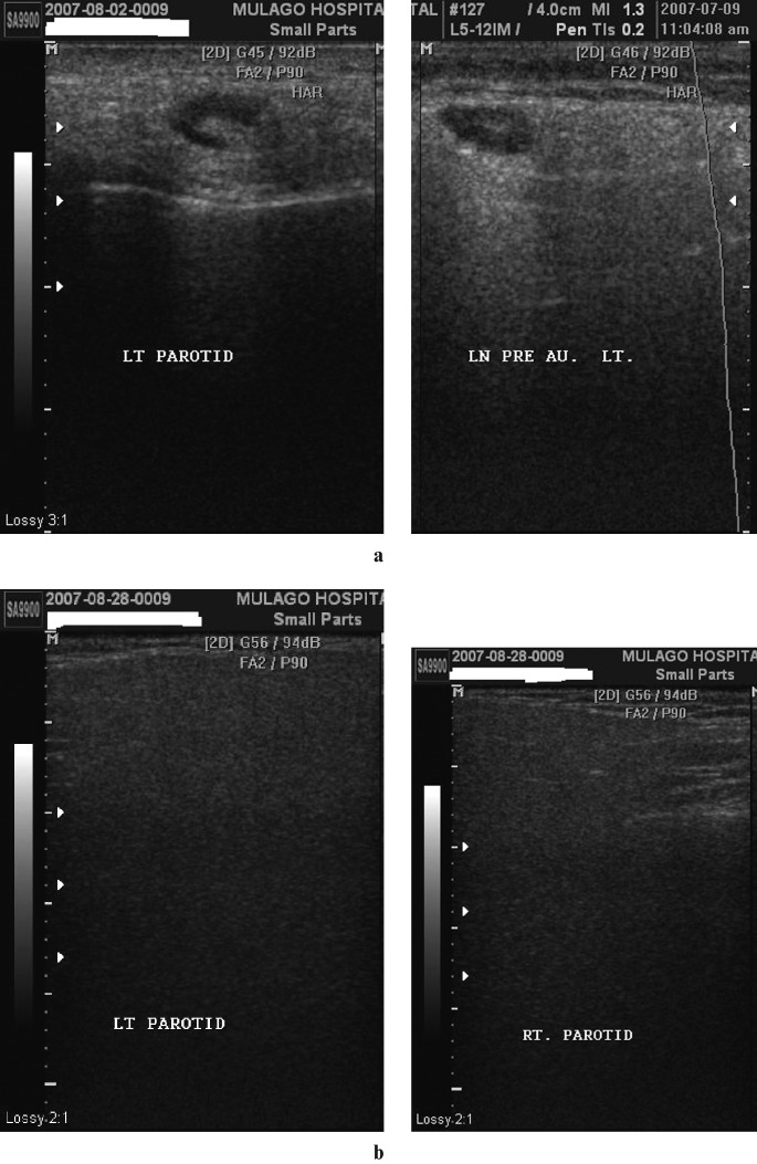
Figure 5.
(a) Fatty infiltration with lymphadenopathy. Coronal sections of a parotid showing an enlarged reactive lymph node within the gland. Note the prominent echogenic hilum and modest posterior acoustic enhancement. (b) Fatty infiltration without lymphadenopathy. Coronal sections of left and right parotids which appear very hypoechoic and homogeneous with posterior attenuation, rendering the images very dark. There are no cysts or lymph nodes

