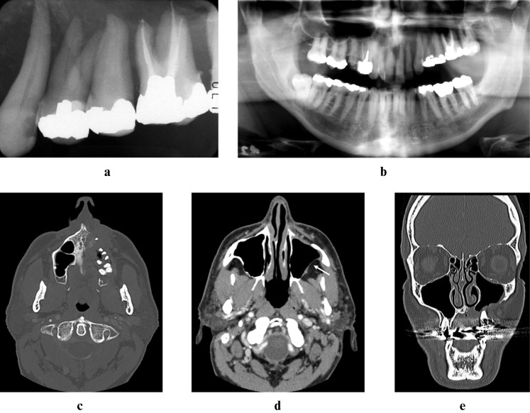
Figure 2.
Digitally enhanced periapical (a), digitally enhanced panoramic (b), axial bone window computed tomography (CT) (c), axial soft tissue-window CT (d) and coronal bone-window CT (e) images at second presentation (2005) demonstrating massive bone destruction of the left maxilla. Despite loss of the cortical boundaries, there is no soft tissue mass present in the maxillary sinus or in the retroantral fat (d, white arrow)

