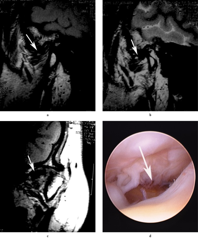
Figure 5.
MRI of a patient (19-year-old woman) with disc perforation, which was confirmed on arthroscopy of the TMJ. (a) Sagittal T1 weighted image in closed-mouth position. (b) Sagittal T1 weighted image in open-mouth position. (c) Coronal T1 weighted image in closed-mouth position revealed anteromedial disc displacement without reduction in the left TMJ (arrows). (d) Disc perforation was confirmed by arthroscopy of the TMJ in the lateral part of the disc (location IIIa)

