Figure 1.
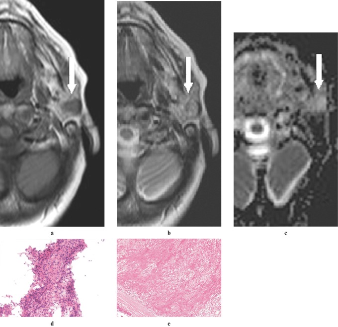
A pleomorphic adenoma in the superficial lobe of the left parotid gland of a 70-year-old woman. (a) An axial T1W image (500/14, TR/TE) showing a mass lesion that is hypointense (arrow) to gland parenchyma and isointense compared with muscle. (b) An axial T2W image (3800/90, TR/TE) showing a mass lesion that is isointense (arrow) with gland parenchyma and hyperintense compared with muscle. (c) An apparent diffusion coefficient (ADC) map showing a solid lesion (arrow) with high ADC (1.54×10⁻³ mm2 s−1). (d) An image of fine-needle aspiration cytology (FNAC) showing spindle-shaped mesenchymal cells intermingled with epithelial cells (haematoxylin and eosin ×20 original magnification). (e) A histological section of the tumour showing biphasic appearance of pleomorphic adenoma resulting from the intimate admixture of epithelium and stroma separated from the surrounding salivary gland tissue by an intact fibrous capsule (haematoxylin and eosin ×10 original magnification)

