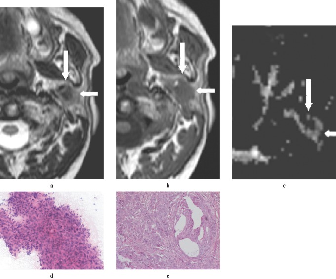
Figure 3.
An adenocarcinoma located in the deep lobe of the left parotid gland of a 62-year-old man. (a) A non-contrast axial T2W image (3800/90, TR/TE) showing a heterogeneous hypointense mass (arrows) relative to the gland parenchyma. Note the ill-defined contour of the lesion. (b) A non-contrast axial T1W image (500/14, TR/TE) showing mass (arrows), hypointense to gland parenchyma. (c) An apparent diffusion coefficient (ADC) map image showing the lesion (arrows) with an intermediate ADC of 1.19 × 10⁻³ mm2 s−1. (d) An image of FNAC showing a cluster of pleomorphic and atypical epithelial cells (hematoxylin and eosin × 20 original magnification). (e) A histological section of the tumour showing adenocarcinoma infiltration in desmoplastic stroma (hematoxylin and eosin × 20 original magnification)

