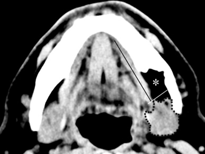
Figure 4.

Mediolateral location of the defect, which consisted of fat tissue of low attenuation (asterisk), submandibular gland (dashed line), mylohyoid muscle (long black line) and distal border of the mylohyoid muscle (short white line) in the axial CT scans using soft-tissue window settings
