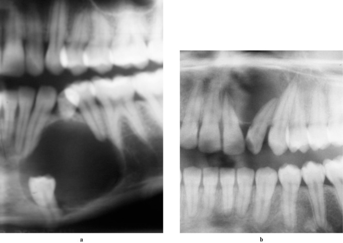
Figure 3.
Radiographs of patients with adenomatoid odontogenic tumours. (a) A cropped panoramic radiograph showing the follicular variety associated with an impacted tooth. (b) A cropped panoramic radiograph of the extrafollicular variety not associated with an impacted tooth. Note the displacement of the lateral incisor and external root resorption of the central incisor

