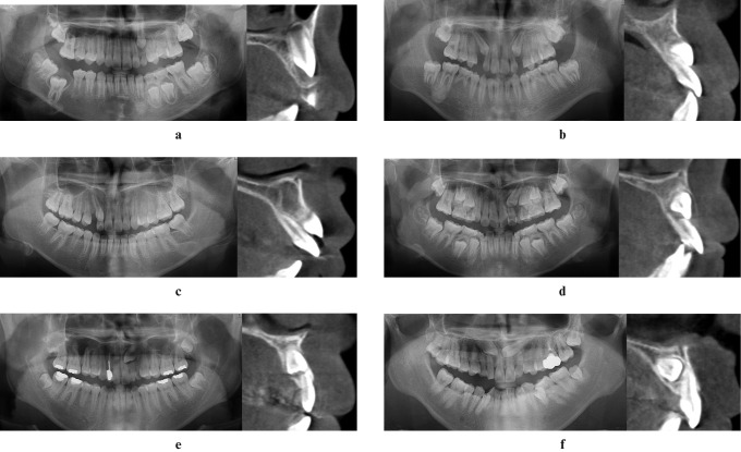
Figure 2.
(a-f) Sector location on panoramic radiograph was compared with the labiopalatal position of impacted maxillary canines in cross-sectional views of cone beam CT images. (a) Left maxillary canine is located in Sector 1 and labially impacted; (b) right maxillary canine is located in Sector 2 and labially impacted; (c) right maxillary canine is located in Sector 3 and labially impacted; (d) left maxillary canine is located in Sector 3 and labially impacted, and lateral incisor shows resorption; (e) left maxillary canine is located in Sector 4 and mid-alveolus impacted, and lateral incisor shows resorption; (f) left maxillary canine is located in Sector 5 and palatally impacted

