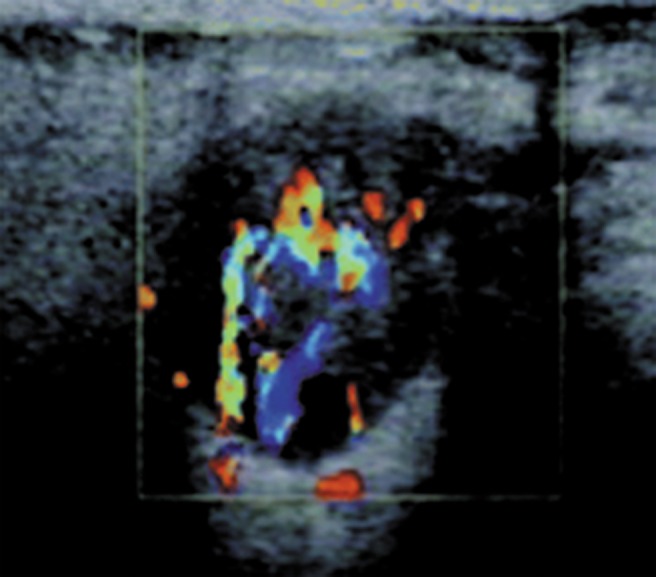
Figure 3.

Sonographic image shows a heterogeneous hypoechoic ovoid mass in the right parotid in a 47-year-old male with circumscribed margin, posterior echogenicity enhancement, distinct edge refraction and much vascularization in colour Doppler imaging. The presumed diagnosis was probably benign lesion and the pathological diagnosis was oncocytoma
