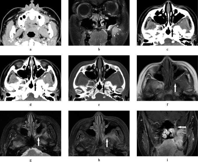
Figure 2.
Adenoid cystic carcinoma of the left cheek in a 27-year-old female (Case 2). (a,b) An ill-defined-margin tumour in the left cheek can be detected on CT and MR images (arrowheads). (c) A plain CT image shows obliteration of fat in the left pterygopalatine fossa (arrow). (d) A contrast-enhanced CT image shows inhomogeneous enhancement in the left pterygopalatine fossa (arrow). (e) A reformatted CT image with bone algorithm shows widening of the left pterygopalatine fossa (arrow). (f) A T1 weighted MR image shows intermediate signal intensity the same as that of adjacent muscle in the left pterygopalatine fossa (arrow). (g) A T2 weighted MR image with the fat suppression (FS) technique shows higher signal intensity than that of adjacent muscle in the left pterygopalatine fossa (arrow). (h,i) A contrast-enhanced T1 weighted MR image with the FS technique shows inhomogeneous enhancement of the left pterygopalatine fossa (arrows)

