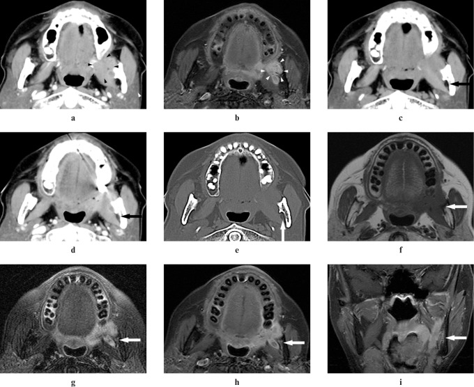
Figure 4.
Adenoid cystic carcinoma of the left retromolar in a 72-year-old female (Case 8). (a,b) An ill-defined margin tumour in the left retromolar can be detected on CT and MR images (arrowheads). (c) A plain CT image does not show elevation of CT value in the left mandibular foramen (arrow). (d) A contrast-enhanced CT image does not show inhomogeneous enhancement in the left mandibular foramen (arrow). (e) A reformatted CT image with bone algorithm does not show widening of the left mandibular foramen (arrow). (f) A T1 weighted MR image shows intermediate signal intensity the same as that of adjacent muscle in the left mandibular foramen and the left mandibular canal (arrow). (g) A T2 weighted MR image with the fat suppression (FS) technique shows higher signal intensity than that of adjacent muscle in the left mandibular foramen and the left mandibular canal and widening of the left mandibular foramen (arrow). (h,i) Contrast-enhanced T1 weighted MR images with the FS technique show contrast enhancement of the left mandibular foramen and the left mandibular canal and widening of the left mandibular foramen (arrows)

