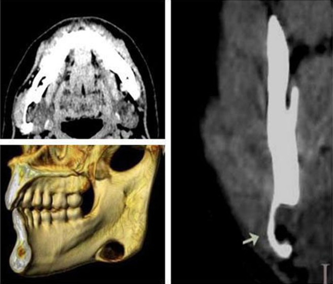
Figure 3.

Case 3. Non-contrast-enhanced CT showing submandibular gland tissue (30 HU). Axial, coronal and three-dimensional CT views show typical apperance of Stafne bone cavity near angle of mandible on right side. The bottom of cavity reaches the buccal cortical plate, which is not expanded (arrow)
