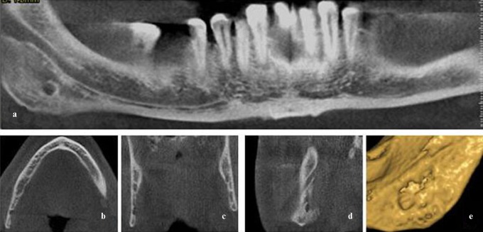
Figure 5.
Case 23. (a) Stafne defect situated in the posterior area of the right mandible below the inferior dental canal. (b) Axial, (c) coronal and (d) sagittal cone beam CT images showing the peripheral origin of the defect and the preservation of the lingual cortex. Bottom of defect does not reach buccal cortical plate. (e) Three-dimensional reconstruction

