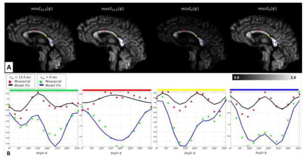
Figure 6.
In vivo detection of microscopic anisotropy using quadruple pulsed-field gradient (qPFG) diffusion MRI on a clinical scanner. A. Extrema (minima and maxima) of angular modulation profiles E13.4(ψ) and E0(ψ) in qPFG diffusion signal attenuation images measured using τm = 13.4 ms and τm = 0 ms, respectively. B. In vivo angular qPFG profiles measured in individual voxels in the prefrontal (green), sensory-motor (red), temporal (yellow) and visual (blue) cortical areas show clear signs of restriction-induced microscopic anisotropy. The modulatory effect of imperfect fiber orientation results in features similar to those in Fig. 2. Nevertheless, theoretical angular qPFG profiles generated numerically using Eq. 4 and high-resolution DTI data reveal good agreement with measurements, validating our methodology for isolating microscopic anisotropy information.

