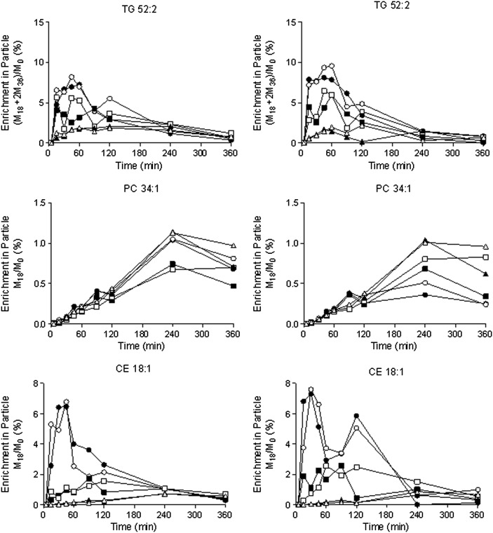
Fig. 3.
Isotopic labeling of different lipids in specific particles. Wild-type mice were given an intravenous injection of [13C18]oleate, and lipoprotein fractions were separated using ultracentrifugation followed by dextran sulfate precipitation (left column) or lipoprint (right column). Data were obtained from 18 different mice (n = 2 per time point). Filled versus open symbols distinguish data from different mice at each time point: VLDL, open/filled circles; LDL, open/filled squares; and HDL, open/filled triangles, respectively. The isotopic labeling of TG, PL, and CE was determined in the various fractions. Data are shown as enrichments, where the notation M18 and M36 refer to the number of labeled oleates that are incorporated; the mass is shifted 18 and 36 units, respectively.

