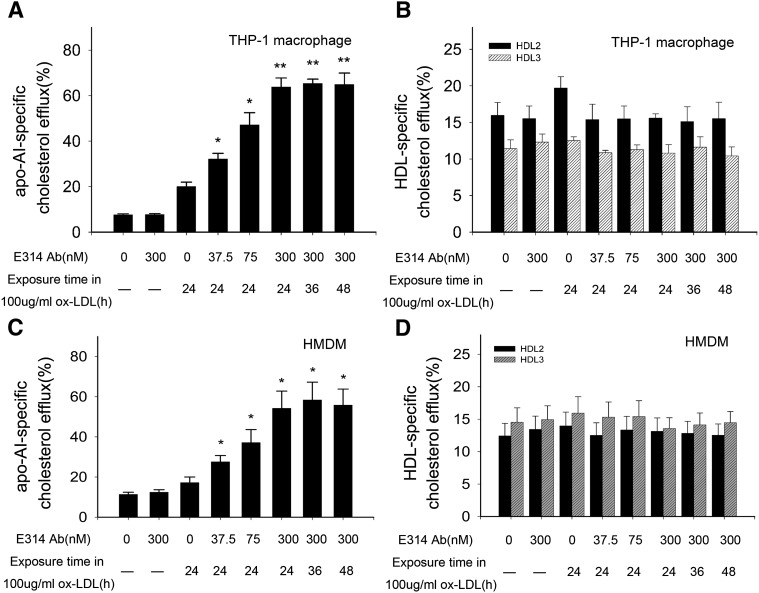
Fig. 2.
Percentage of cholesterol efflux from THP-1 macrophages or HMDMs exposed to 100 μg/ml ox-LDL in the absence (macrophages and macrophages exposed to 100 μg/ml ox-LDL or the 300 nM hKv1.3-E314 antibody alone) or presence of the hKv1.3-E314 antibody at varying concentrations of 37.5, 75, or 300 nM. In the presence of the 300 nM hKv1.3-E314 antibody, macrophages were exposed to 100 μg/ml ox-LDL for 24, 36, or 48 h. Medium and cell-associated apoA-I or HDL-mediated [3H]cholesterol were measured by liquid scintillation counting (A–D). A and C: Percentage of apoA-I-mediated cholesterol efflux in THP-1 macrophages and HMDMs,. B and D: Percentage of HDL-mediated cholesterol efflux in THP-1 macrophages and HMDM cells (n = 3). *P < 0.05, **P < 0.01, and P > 0.05 versus macrophages exposed to 100 μg/ml ox-LDL alone. There was no significant difference in percentage of apoA-I or HDL-mediated cholesterol efflux when macrophages were exposed to 100 μg/ml ox-LDL for 24, 36, or 48 h in the presence of the 300 nM hKv1.3-E314 antibody.

