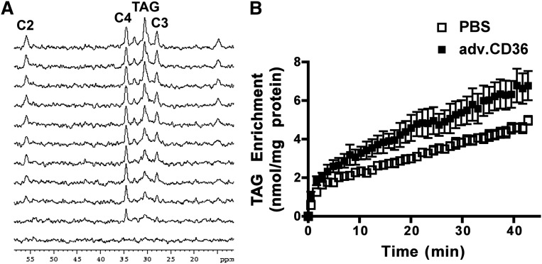
Fig. 3.
Incorporation of 13C-palmitate into intramyocardial TAG. (A) Representative 13C spectra from a control intact perfused heart obtained sequentially (1 min acquisition, from bottom). For simplicity of presentation, only every fourth trace is presented. (B) 13C enrichment of the TAG pool as a function of time was determined from signal intensity of the methylene at 30.5 ppm. Data are presented as means ± SE. n = 5 for sham and adv.CD36.

