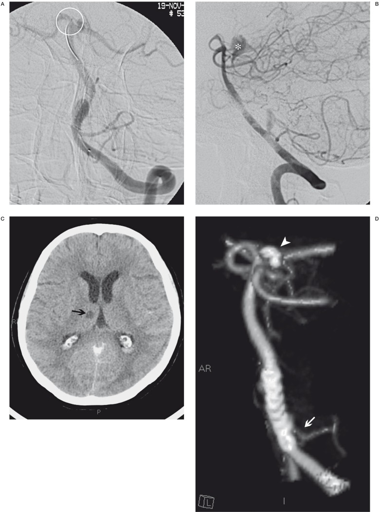
Figure 2.
A) The position where the guide tip was accidentally blocked during stent release (ring). When we performed a final angiographic control (B) we observed a leakage of contrast into the interpeduncular cistern (*). Two days later, CT control (C) disclosed a right thalamic ischaemic lesion (black arrow) and subtotal reabsorption of leakage of contrast. Two months later, CT angiography follow-up control showed the aneurysm occlusion, regular view of the PICA (white arrow), good position of the Silk stent and presence of the coils (arrowhead) that we used to block the haemorrhage due to the artery of Percheron perforation.

