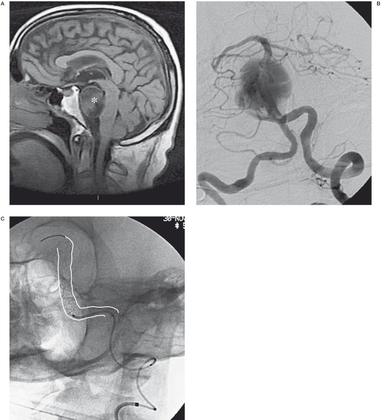
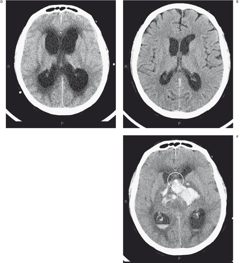
Figure 5.
A-C) The MR image shows a giant basilar aneurysm and its massive compression on the bulb (*). Angiographic examination confirms the giant aneurysm that was treated with one Silk stent (white lines). D-F) CT controls post endovascular treatment showed acute hydrocephalus that was treated by external CSF drainage (ring), but it resulted in massive intra-axial and intraventricular haemorrhage.


