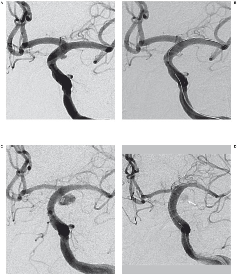
Figure 3.
A 52-year-old man (patient No. 3) presenting with HH grade IV SAH. A) Preprocedural DSA shows a blister aneurysm along the lateral wall of the supraclinoid segment of left ICA. B) A control angiogram obtained after stent-assisted coil embolization, revealed minimal residual aneurismal filling. C) Follow-up left ICA angiogram obtained 12 days postoperatively revealed considerable BBA growth. D) Immediately after re-treatment with a covered stent, the control angiogram revealed subtle contrast media leakage into the recurred sac (arrow).

