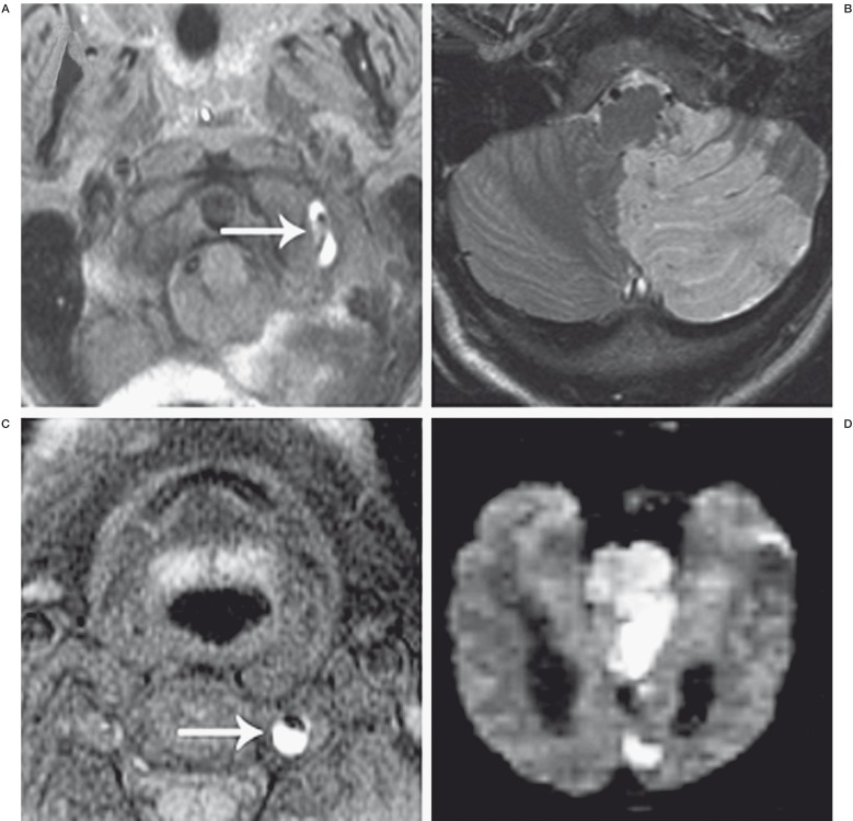
Figure 1.
Ischemic complications of vertebral artery dissection. A 46-year-old woman presented with worsening headaches, nausea, and dizziness one week after the acute onset of left-sided neck pain during chiropractic manipulation. A) Axial T1 fat saturation MRI demonstrates a hyperintense signal surrounding the left vertebral artery (white arrow) indicating dissection. B) Axial FLAIR MRI shows a subacute infarct in the distribution of the left PICA. C) Axial T1 fat saturation MRI in a 32-year-old man who suffered the acute onset of left-sided neck pain without preceding trauma. The left vertebral artery is shown to be dissected, as evidenced by the hyperintense intimal flap and associated thrombus (white arrow). D) Diffusion-weighted MRI demonstrates diffuse acute infarcts in the pons, left occipital lobe, and left mesial temporal lobe. The patient presented intubated and was found to be locked-in on examination.

