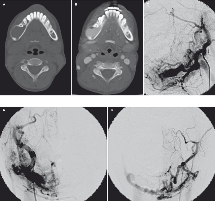
Figure 1.
Axial CT images of head and neck without (A) and with (B) i.v. contrast medium enhancement showed an intraosseous lytic expansile lesion with avid enhancement at body and ramus of the right mandible. The right carotid angiogram in lateral (C) and AP (D) projection and left carotid angiogram in AP projection (E) demonstrated complex intraosseous and extraosseous AMV in right mandible and pterygoid region. The AVMs was supplied from bilateral ECAs and drained mainly by the right retromandibular vein.

