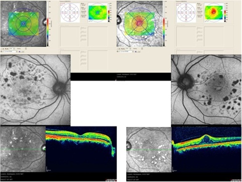
Figure 5.
Two eyes with diabetic macular edema which have received macular photocoagulation; the last row shows optical coherence tomography images of the two eyes and the middle row shows autofluorescence imaging. In the right eye (left image), macular edema has resolved and there is no hyper-autofluorescence in the macular region. In the left eye (right image) there is persistent macular edema and visible hyper-autofluorescence in the macula.

