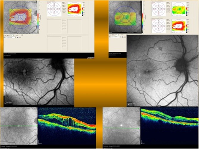
Figure 6.
The amount of macular edema is more evident on images in the left column (baseline macular edema) as compared to right side images (after treatment). Left side fundus autofluorescence shows more hyperautofluorescence in the foveal center. Although there is retinal edema on the temporal side of the fovea, hyper-autofluorescent is not seen in the parafoveal area.

