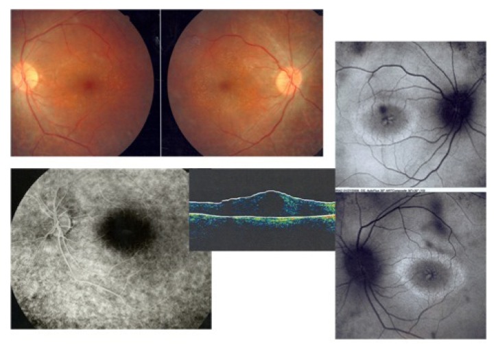
Figure 7.
Retinitis pigmentosa and cystoid macular edema; fundus photograph images of both eyes show typical arteriolar narrowing and bony spicules (upper left images), fluorescein angiography shows no leakage in the macular area (lower image), autofluorescence images of both eyes reveals hyperfluorescent cysts corresponding to cystic edema (right side images), besides a hyperautofluorescent ring in the macula shows the border between involved and uninvolved photoreceptors. The ring becomes smaller with disease progression.

