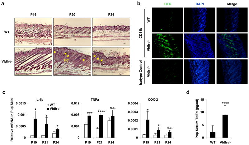
Figure 2. Maternal VLDLR deletion causes neonatal inflammation.
a–c, Defects in the skin of the pups nursed by Vldlr−/− mothers compared with the pups nursed by WT control mothers. a, H&E staining showed skin hyperplasia and follicular cysts (FC, indicated by arrows). Scale bars, 100μm. b, Immuno-fluorescence staining showed leukocyte infiltration in the skin at P19. Skin sections were stained with a FITC-CD11b antibody or FITC-isotype control (to show non-specific staining of the hair shafts). Scale bars, 100μm. c, RT-QPCR analysis showed increased inflammatory gene expression in the skin (n=3). d, Serum level of TNFα was elevated in the pups nursed by Vldlr−/− mothers compared to the pups nursed by WT control mothers (n=12, P19). The pups nursed by Vldlr−/− mothers were Vldlr+/−, and the pups nursed by WT mothers were WT. Statistical analyses were performed with Student’s t-Test and are shown as mean ± standard deviation; *, p<0.05; ***, p<0.005; ****, p<0.001; n.s., non-significant.

