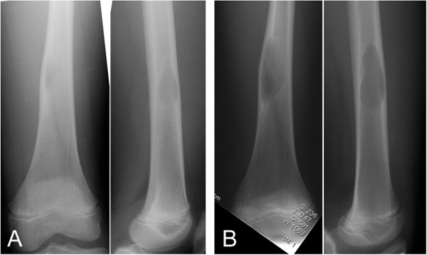
Figure 1.

Antero-posterior (left) and lateral (right) plain radiographs show a well-defined osteolytic lesion in the medullary space with cortical thinning (A). The size of the lesion increased over 10 months after the initiation assessment on antero-posterior (left) and lateral (right) views (B).
