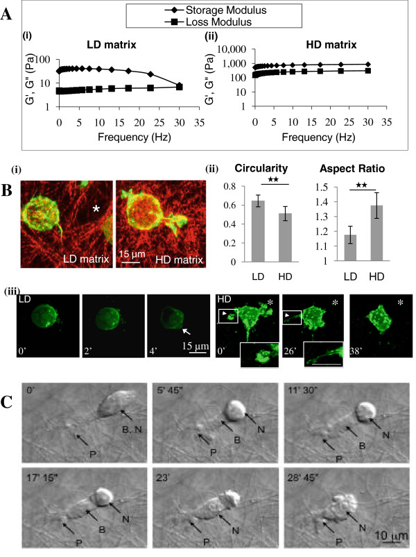
Figure 2.
Matrix properties and cell migration. Tumour cells were grown in low-density (LD) matrix of 1 mg/cm3 or high-density (HD) matrix of 20 mg/cm3. A, The storage modulus (G’, ♦) and the loss modulus (G”, ■) of matrices were graphed against oscillation frequency sweeping from 0.01 to 30 Hz. As the rate of deformation increases, the storage modulus and loss modulus values converge at high deformation rates in LD but not HD matrix reflecting higher viscoelastic properties in the latter. B, (i), Merged reflection and confocal images of collagen and paxillin-Alexa 568-labelled tumour cells demonstrate larger interfibrillar spaces (*) in LD matrix. (ii) Morphometric indicators show that tumour cells are more rounded in shape in LD compared to HD matrices (n = 15). (iii), Live-cell confocal microscopy of GFP-actin transfected MTLn3 cells. Cells in LD matrix appear rounded and form blebs during migration (arrow). In HD matrix, protrusions are extended via ruffling (arrow heads) and terminate as fine filopodia (inset images). Asterisk indicates the initial cell position. C, Live-cell DIC microscopy shows a typical migration pattern of a tumour cell through HD matrix. At time 0’, a cell protrusion (P) has breached the fibrous mass. At 5’ 45”, the cell body (B) has partially extended into the matrix via cytoplasmic propulsion leaving behind the rounded, tall nucleus (N). In later frames, contraction of the cell body facilitated squeezing of the nucleus past obstructing matrix fibrils, completing the migration cycle by 28’ 45”.

