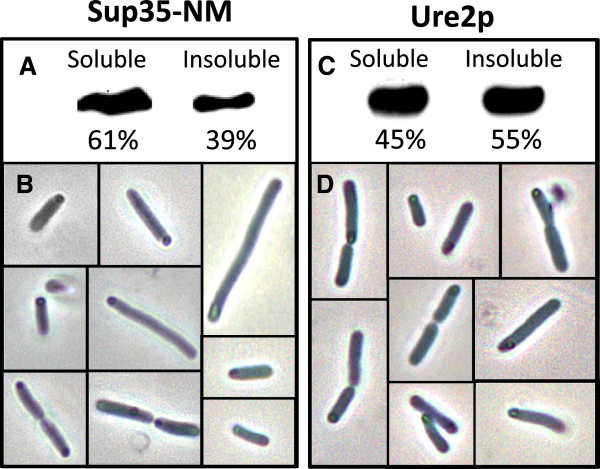
Figure 1.
Solubility properties of recombinant Sup35-NM (left panel) and Ure2p (right panel) proteins. (A and C) Western blot of the soluble and insoluble fractions of cells expressing Sup35-NM and Ure2p at 37°C detected with an anti-histag antibody and quantified by densitometry using the Quantity-One software (Bio-Rad). (B and D) Localization of cytoplasmic IBs at the poles of cells expressing Sup35-NM and Ure2p proteins, as imaged by phase contrast microscopy.

