Figure 4.
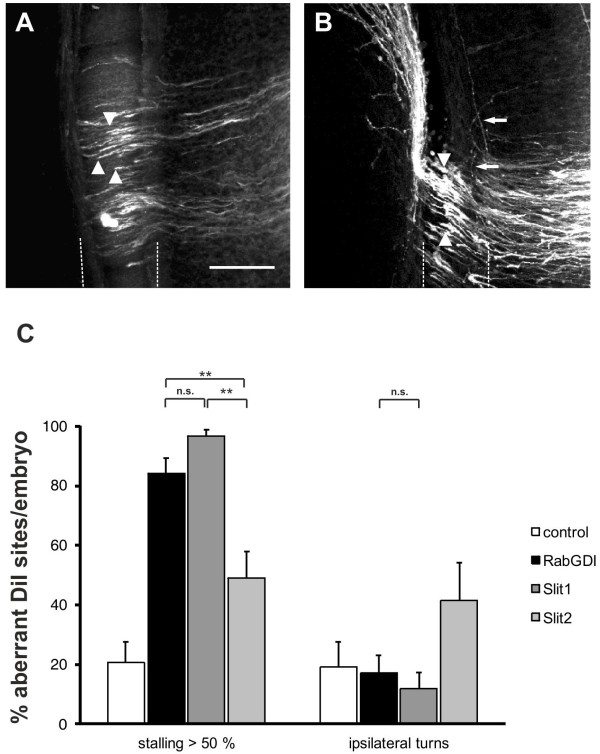
The reduction of negative cues associated with the floor plate by downregulation of Slit1 reproduces the RabGDI phenotype. Silencing Slit1 (A) or Slit2 (B) by in ovo RNAi resulted in stalling of commissural axons (arrowheads) in the floor plate (dashed lines), similar to the phenotype observed after loss of RabGDI function (compare to Figure 1C and D). However, the effect of loss of Slit2 on commissural axon stalling was significantly weaker than the effect of Slit1 downregulation (C). Loss of Slit1 or Slit2 function also revealed qualitative phenotypic differences between the two. The Slit1 phenotype was the same as the one seen in the absence of RabGDI (compare A with Figure 1C, D). In contrast, loss of Slit2 additionally produced axons that turned into the longitudinal axis prematurely (arrows in B), an abnormality that was only rarely seen after downregulation of Slit1 (C). Phenotype quantifications are shown in (C). Significance levels: n.s. not significant, ** p<0.01. Values for control and RabGDI taken from Figure 2C. Bar: 50 μm.

