
Figure 2.
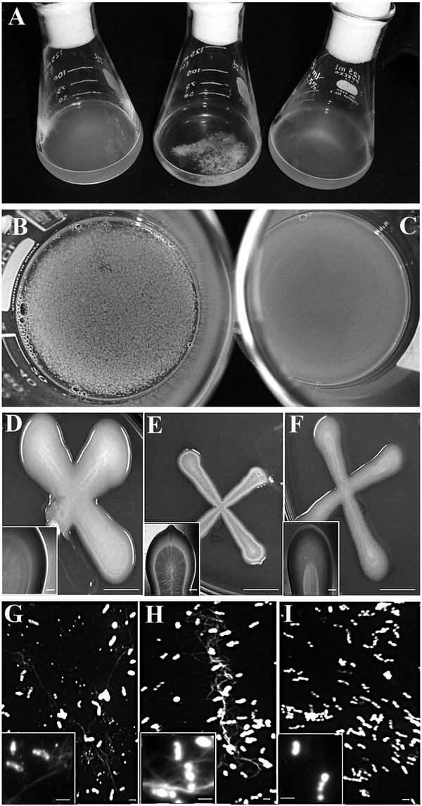
From left to right, strain ANU843 (wild type) and its derivatives ANU843ΔC2 and ANU843C2+. (A) Batch cultures in stationary phase in YMB shaken at 180 rpm. (B-C) ANU843ΔC2 flocs after incubation 2h at 37°C in PCA buffer pH 5 (B) and containing 10 U/ml Trichoderma viride commercial cellulase (C). (D) Line streak colonies grown on TY (bar = 1cm). The insert in the lower left corner is an enlargement of the images (bar = 1 mm). (G-I) Calcofluor staining showed the presence of micro fibrils (bar = 1.0 μm) in wild type ANU843 (G), ANU843ΔC2 (H) and ANU843C2+ (I). Inserts at the lower left corners show representative cells at higher magnification. In the insert images note the accumulation of bright fluorescent target at both cell poles separated by reduced fluorescence intensity midway between the cell poles.