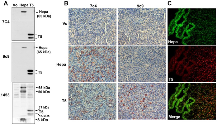
Figure 1. Characterization of anti T5 mAb.
A. Immunoblotting. HEK 293 cells were transfected with heparanase (Hepa) or T5 gene constructs and lysate samples were subjected to immunoblotting applying mAb 7c4 (upper panel), mAb 9c9 (middle panel) or pAb 1453 (lower panel). Cells transfected with an empty vector (Vo) were used as control. Note that mAb 9c9 preferentially recognizes T5 vs. heparanase. B. Immunohistochemistry. Five micron sections of tumor xenografts produced by control CAG myeloma cells (Vo) or CAG cells over expressing heparanase (Hepa) or T5 were subjected to immunostaining applying mAb 9c9 as described under 'Materials and Methods'. Note that mAb 9c9 only reacts on sections derived from CAG cells over expressing T5. C. Human placenta. Placenta specimens were subjected to double immunofluorescent staining applying rabbit (pAb 733, green) and mouse (mAb 9c9, red) anti-heparanase antibodies. Merged image is shown in the lower panel. Note distinct expression pattern of heparanase (cytotrophoblasts) and T5 (villus stromal cells).

