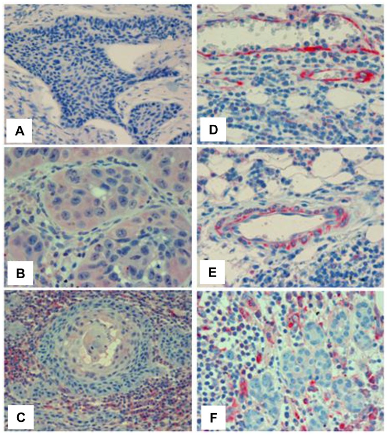
Figure 2. T5 staining in head and neck tumor biopsies.
Tumor specimens were subjected to immunohistochemical analyses applying mAb 9c9 as described under 'Materials and Methods'. Shown are representative images of head and neck T5 negative (A) and positive tumors expressing T5 in tumor cells (B), tumor microenvironment (C, F), and tumor-associated blood vessels (D, E).

